3D Computer-Guided SurgeryCASE-06Verified Clinical Registry · 25x Carl Zeiss
Computer-Guided 3D Precision Implant Placement with Surgical Template
100% pre-operative digital simulation and 3D printed surgical stent ensuring sub-millimeter precision and flapless placement.
Interactive Visual Comparison
Drag slider left/right or use arrow keys to inspect the transition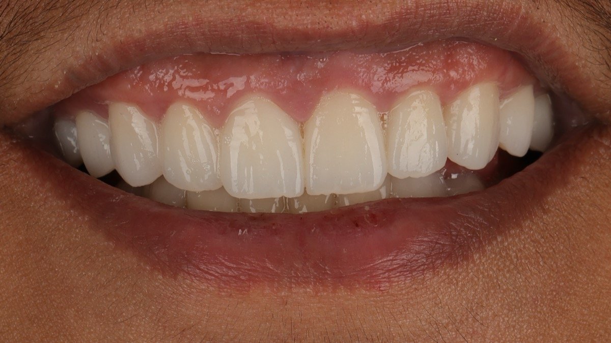
Final Result
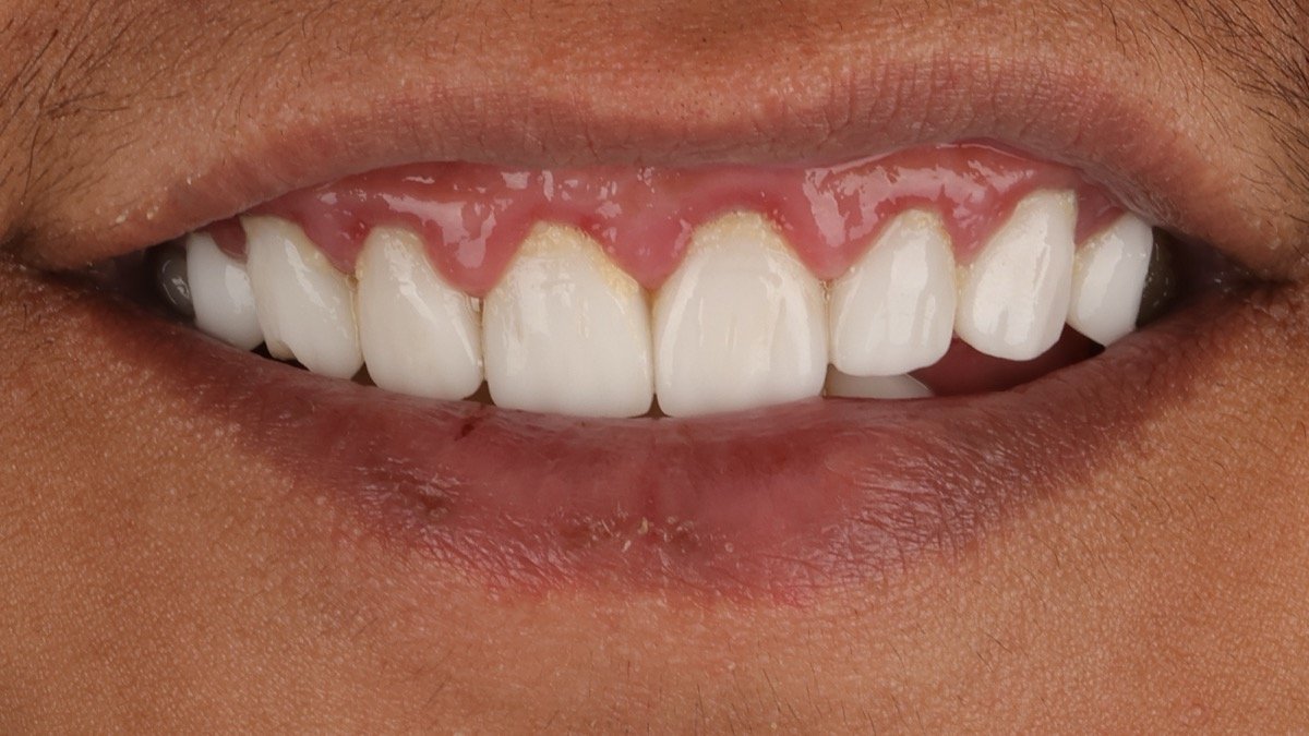
Before Treatment
* High-resolution intraoral clinical photography documented under surgical illumination.Published with written patient consent
Procedural Specifications & Clinical Metrics
Technology3D Printed Surgical Stent
InvasivenessFlapless Micro-Invasive
SafetyZero Nerve Risk
Clinical Report & Diagnostic Protocol
TRIOS intraoral optical scans were married with 3D CBCT datasets to calibrate the exact osteotomy trajectory. Implants were placed through a 3D-printed stereolithographic guide with minimal soft-tissue disruption and zero scalpel flaps.
Radiographic & 3D CBCT Validation
Pre-operative and post-operative radiographic verification demonstrating osseointegration, cortical bone density, and precise anatomical clearance.
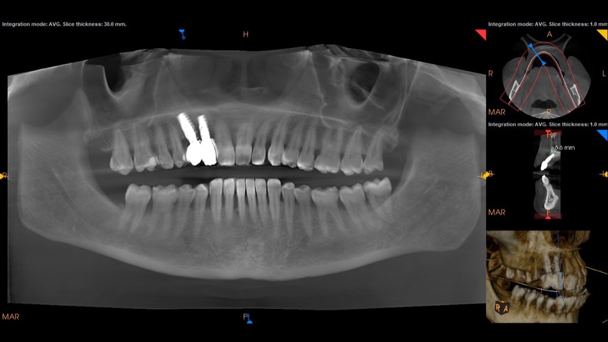
Diagnostic Scan #1Validated
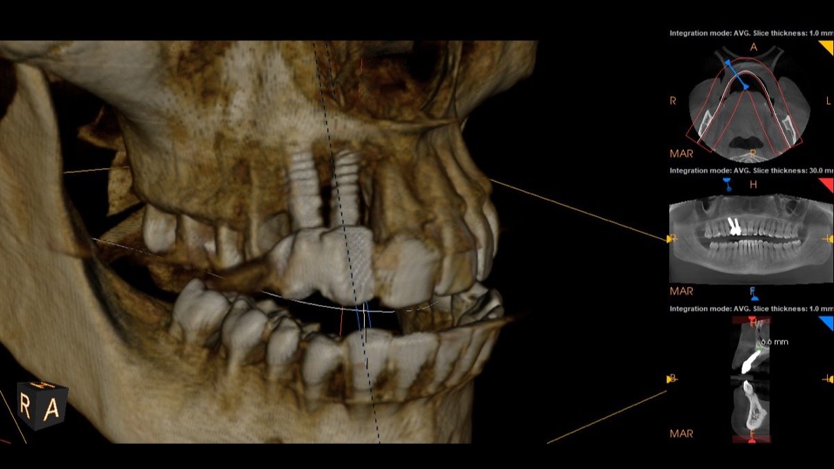
Diagnostic Scan #2Validated
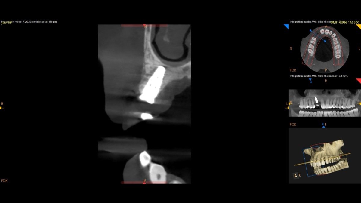
Diagnostic Scan #3Validated
Surgical & Restorative Technologies Applied
3Shape TRIOSCBCT Guide CalibrationFlapless Surgery
Personalized Case Assessment
Presenting a Similar Clinical Condition?
Send your panoramic X-ray or intraoral photographs directly to Dr. Shadi Alshaikh for a preliminary assessment and treatment feasibility plan.